Haiye Medical Haiye Medical

Top 10 Fiber Optic Laryngoscope Uses You Should Know?

Time:2026-09-18 Author:Mason
0%

A fiber optic laryngoscope is more than a viewing instrument. It is a slender, illuminated guide that helps clinicians examine hidden airway structures with greater control. Its flexible tip can pass around anatomical curves, while the camera displays the epiglottis, vocal cords, and nearby tissues on a monitor. These details matter in operating rooms, emergency departments, intensive care units, and specialist clinics.

The most important fiber optic laryngoscope uses include difficult-airway assessment, guided tracheal intubation, vocal-cord examination, foreign-body evaluation, biopsy assistance, and postoperative airway review. It can also support teaching, documentation, and careful follow-up. Dr. John D. Ovassapian, a respected authority in fiber-optic airway management, emphasized, “Fiberoptic intubation is a skill that requires practice, patience, and preparation.” That principle remains highly relevant. Equipment alone cannot replace clinical judgment.

A quiet room helps.

The image may reveal swelling, secretions, bleeding, or unexpected anatomy. Yet the view is not always perfect. Fogging, limited visibility, patient discomfort, and operator inexperience can affect results. This is where many simplified explanations fall short. The device does not automatically create a safe airway. Trained professionals must select the correct technique, monitor the patient continuously, and follow local clinical protocols.

This guide explores the top 10 fiber optic laryngoscope uses through practical examples. It considers where the instrument helps, what clinicians look for, and which limitations deserve attention. Readers should also remember that device design, training standards, and clinical indications vary between healthcare settings.
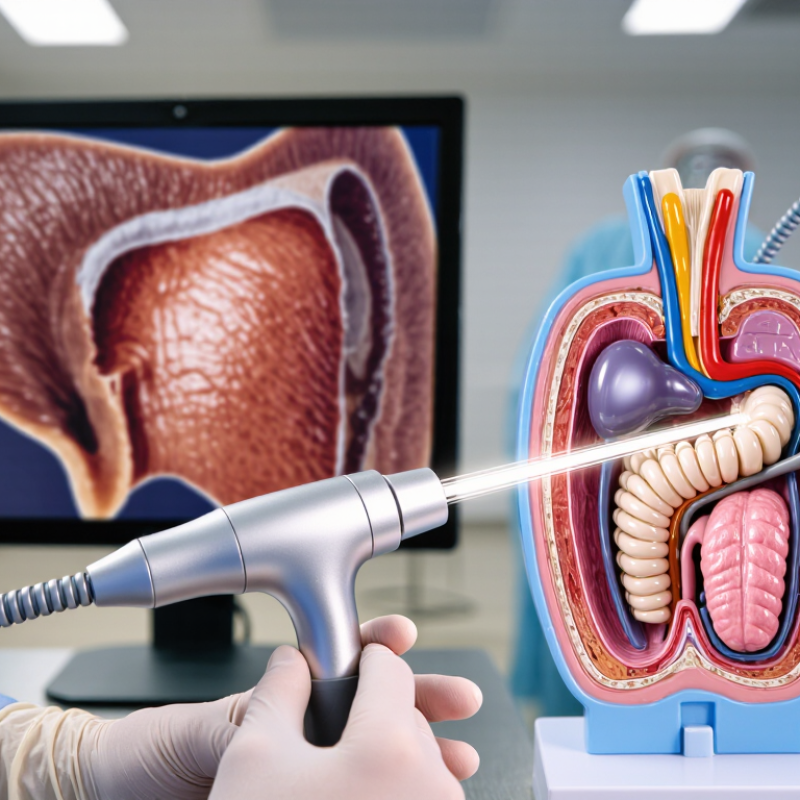
Top 10 Fiber Optic Laryngoscope Uses You Should Know?

What Is a Fiber Optic Laryngoscope and How Does It Work?

A fiber optic laryngoscope is a slender, flexible instrument for viewing the nose, throat, and larynx. It uses fiber bundles or a miniature camera to transmit images to an eyepiece or monitor. A clinician guides its curved tip around the tongue and epiglottis. The vocal cords then appear as moving white bands during breathing and speech.

Its uses include airway assessment, vocal-cord examination, difficult-airway evaluation, awake intubation, post-operative review, swallowing assessment, voice analysis, foreign-body investigation, and selected tissue procedures. The Fourth National Audit Project (NAP4) reported one major airway complication for approximately every 22,000 general anesthetics. This supports careful airway planning, although the figure does not measure laryngoscope performance directly. The 2022 American Society of Anesthesiologists guidelines recognize flexible intubation as an important option when difficult airway management is anticipated.

Tips: Use topical anesthesia only under trained clinical supervision. Keep the lens clear, control secretions, and avoid forcing the tip. A blurred image can hide swelling or bleeding. That risk is easy to underestimate. Fiber optics also demand practice; hand movement, patient anatomy, and lighting affect image quality. In my view, the device is powerful but not magical. A backup airway plan remains essential, especially when oxygen levels fall quickly or the patient cannot cooperate.

Top Diagnostic Uses of Fiber Optic Laryngoscopy

Top 10 Fiber Optic Laryngoscope Uses You Should Know?
Top Diagnostic Uses of Fiber Optic Laryngoscopy

Fiber optic laryngoscopy gives clinicians a close view of the throat and voice box. A thin flexible scope usually passes gently through one nostril. A camera then displays the vocal folds on a monitor. This examination can investigate persistent hoarseness, weak voice, throat discomfort, and unexplained coughing. It can also reveal vocal fold nodules, polyps, swelling, ulcers, or suspicious tissue changes. The view may show vocal fold paralysis or unequal movement during speech. That detail often helps guide further testing.

This procedure also supports swallowing assessments. Clinicians can observe secretions, delayed movement, and possible aspiration around the larynx. It may help identify airway narrowing, irritation linked with reflux, or changes after surgery. In children and adults, it can assist with evaluating noisy breathing when appropriate. The examination is quick, but not effortless. The view can be blurred by mucus, movement, or a strong gag reflex. Findings require professional interpretation, and some lesions still need imaging or tissue sampling. Patient comfort, informed consent, and careful infection control remain essential. A normal-looking examination does not explain every symptom. Persistent or worsening problems may require repeated assessment.

Top 10 Fiber Optic Laryngoscope Uses You Should Know

Fiber optic laryngoscopy provides direct visualization of the nasal passages, pharynx, larynx, vocal folds, and upper airway. The chart groups ten established diagnostic applications by their primary clinical purpose. The values represent the number of applications in this summary, not procedure frequency.

Diagnostic Uses Included

  • Assessment of vocal fold movement and symmetry
  • Detection of vocal fold nodules, polyps, cysts, and other lesions
  • Evaluation of glottic closure during phonation
  • Identification of laryngeal inflammation and edema
  • Inspection of supraglottic abnormalities
  • Evaluation of vocal fold paralysis or paresis
  • Detection of hypopharyngeal lesions
  • Assessment of swallowing safety and aspiration risk through flexible endoscopic evaluation
  • Investigation of upper-airway narrowing or obstruction
  • Follow-up surveillance after treatment for laryngeal disease

Clinical note: Flexible laryngoscopy is commonly used to investigate hoarseness, voice changes, swallowing complaints, suspected airway problems, and visible abnormalities of the upper aerodigestive tract. The exact examination depends on the patient’s symptoms and the clinician’s assessment.

Top Clinical Procedures Performed with a Fiber Optic Laryngoscope

A fiber optic laryngoscope supports several important clinical procedures, especially when direct examination is difficult. Flexible transnasal laryngoscopy allows clinicians to inspect the nasal passages, pharynx, epiglottis, and vocal folds. It is commonly used for hoarseness, throat discomfort, chronic cough, swallowing problems, and unexplained noisy breathing. The patient remains awake, which helps clinicians assess natural speech and vocal fold movement.

Airway assessment is another major use. During difficult intubation, the flexible scope can guide the tube while providing a continuous view of the airway. It may also help evaluate narrowing, swelling, secretions, or unexpected anatomy before surgery. After an operation, clinicians can check vocal fold mobility and identify possible irritation or injury. The procedure requires careful preparation, topical anesthesia, infection control, and informed consent.

The view is not always perfect. Blood, mucus, patient movement, or a sensitive gag reflex can limit examination quality. Experienced clinicians may need to pause, reposition the scope, or repeat the assessment. A fiber optic laryngoscope can reveal useful findings, but it does not replace a complete medical history or other tests. Any suspicious lesion may require further evaluation, imaging, or tissue sampling by a qualified specialist.

Emergency, Airway Management, and Pediatric Applications

Top 10 Fiber Optic Laryngoscope Uses You Should Know?
Emergency, Airway Management, and Pediatric Applications

In emergency care, a flexible fiber optic laryngoscope can guide nasal or oral intubation when direct views fail. It supports anticipated difficult airways, restricted mouth opening, facial trauma, cervical spine precautions, and tube exchange. Real-time visualization helps clinicians navigate around the tongue and epiglottis while limiting neck movement. The 2022 American Society of Anesthesiologists guidelines recommend considering awake intubation when difficult ventilation, aspiration risk, apnea intolerance, or emergency invasive access is expected. NAP4 reported major airway complications at about 1 in 22,000 anesthetics. That figure is a warning, not reassurance.

Pediatric use demands smaller equipment, steady oxygenation, and short attempt times. The Pediatric Difficult Intubation Registry recorded 1,838 difficult pediatric cases across 13 hospitals, showing why rescue planning cannot depend on one device. Fiber optic scopes support anticipated syndromic airways, craniofacial abnormalities, limited neck motion, and teaching with recorded airway views. They can also assess laryngeal anatomy, assist bronchial blocker placement, and help verify tube position when anatomy is uncertain. Yet blood, secretions, fogging, and movement can erase the image quickly. The scope is not magic. Teams need suction, oxygen delivery, backup laryngoscopy, and a clear failure threshold. This point is often under-practiced. Regular simulation and documented competency remain essential, especially for children. Sources: American Society of Anesthesiologists, 2022; Fourth National Audit Project, 2011; Pediatric Difficult Intubation Registry publications.

Safety, Preparation, Limitations, and Equipment Selection

Top 10 Fiber Optic Laryngoscope Uses You Should Know?

A fiber optic laryngoscope can support difficult airway assessment, awake intubation, vocal cord inspection, and postoperative checks. It may also help locate secretions, evaluate throat injuries, guide tube placement, and investigate persistent hoarseness. Trained clinicians should perform these procedures with continuous oxygen monitoring. Safety depends on gentle handling, adequate suction, and a prepared rescue airway plan. A clean lens does not guarantee a clear view.

Preparation begins with a focused history, consent, allergy review, and airway examination. Check the light source, camera, angulation, suction, and recording function before use. Keep oxygen, monitoring equipment, and alternative airway devices nearby. Topical anesthetic requires careful dosing and documentation. Sedation can reduce cooperation, but excessive sedation may weaken spontaneous breathing. That risk is easy to underestimate.

Limitations include blood, mucus, fogging, narrow anatomy, and restricted movement. The image can look acceptable while depth remains difficult to judge. I still recheck positioning rather than trusting the screen alone. Equipment selection should match the patient’s age, anatomy, and procedure. Consider insertion diameter, image quality, tip control, working channels, durability, and validated cleaning methods. Flexible systems suit curved pathways, while rigid devices may provide stronger alignment in selected cases. Reprocessing instructions must be followed precisely, because hidden contamination can remain after visual cleaning.

Top 10 Fiber Optic Laryngoscope Uses You Should Know? - Safety, Preparation, Limitations, and Equipment Selection

No. Common Use Typical Clinical Purpose Preparation Key Safety Measures Important Limitations Equipment Selection Considerations
1 Awake Flexible Intubation Provides a controlled view of the airway when difficult intubation is anticipated or when maintaining spontaneous breathing is desirable. Review the airway, explain the procedure, apply monitoring, prepare suction, and use topical airway anesthesia according to local protocol. Maintain oxygenation, use careful topicalization, avoid excessive sedation, and keep a backup airway plan immediately available. Blood, secretions, edema, patient intolerance, or limited operator experience can obscure the view or prolong the procedure. Choose a maneuverable, appropriately sized scope with a working channel when suction or tube guidance may be needed.
2 Assessment of Vocal Fold Motion Examines vocal fold abduction and adduction in patients with hoarseness, suspected paresis, or possible recurrent laryngeal nerve dysfunction. Obtain symptom history, inspect the nose and oropharynx, and use topical anesthetic only when clinically appropriate. Advance gently under direct visualization and avoid unnecessary contact with the laryngeal structures. It may not identify the cause of impaired motion and may be affected by pain, anxiety, poor cooperation, or inadequate visualization. A high-quality image system, adequate illumination, and recording capability are useful for comparison and documentation.
3 Evaluation of Hoarseness Visualizes the larynx to assess inflammation, lesions, structural changes, or other possible causes of persistent voice symptoms. Confirm the duration and risk factors of symptoms, explain possible discomfort, and prepare a clean examination area. Use gentle insertion, avoid force, and stop if significant pain, bleeding, respiratory distress, or vasovagal symptoms occur. A flexible examination may not replace stroboscopy, imaging, biopsy, or specialist assessment when a detailed tissue evaluation is required. Select equipment with clear visualization, reliable white balance, image capture, and a comfortable distal tip diameter.
4 Investigation of Stridor Helps identify upper-airway narrowing, vocal fold dysfunction, supraglottic collapse, or other causes of noisy breathing. Assess respiratory stability first, prepare oxygen and suction, and ensure personnel capable of airway rescue are available. Do not delay emergency airway management; perform the examination only when the patient is adequately supported and monitored. Instrumentation can worsen distress or obstruction, and a normal examination at one moment may not exclude intermittent problems. Use a small, highly maneuverable scope with dependable illumination and rapid connection to a display.
5 Post-Extubation Airway Review Assesses vocal fold movement, laryngeal swelling, secretions, or structural injury after removal of an endotracheal tube. Confirm clinical stability, prepare oxygen and suction, and coordinate the examination with the airway and critical-care team. Monitor continuously and be ready to provide supplemental oxygen, reintubation, or alternative airway support if deterioration occurs. Findings can change quickly and may not predict delayed airway swelling or respiratory failure in every patient. A portable system with rapid startup, strong suction compatibility, and easy cleaning is advantageous in critical-care settings.
6 Foreign-Body Assessment Supports inspection of the upper airway when aspiration or an obstructing object is suspected. Evaluate airway severity, keep suction and emergency equipment ready, and maintain a low threshold for specialist intervention. Avoid pushing the object deeper; severe obstruction requires immediate emergency management rather than a prolonged diagnostic examination. The scope may not retrieve the object, control bleeding, or adequately examine lower airways; rigid endoscopy may be necessary. Consider a working channel, effective suction, sufficient insertion length, and compatibility with retrieval accessories when indicated.
7 Airway Examination Before Surgery Documents airway anatomy and vocal fold function before procedures that may affect the larynx, neck, or recurrent laryngeal nerves. Record baseline symptoms and findings, verify consent, and document images or video according to institutional policy. Use standard infection-control procedures and avoid unnecessary instrumentation in patients with unstable respiratory status. A baseline examination cannot eliminate all perioperative airway risks and may not detect lesions outside the field of view. Select equipment with consistent image quality, date-stamped recording, and secure data handling for longitudinal comparison.
8 Guidance During Airway Procedures Provides visual guidance for tube placement, airway exchange, or confirmation of endotracheal tube position when direct visualization is difficult. Check the scope, light source, suction, tube compatibility, oxygen delivery, and backup airway equipment before starting. Confirm tube position with clinical assessment and appropriate monitoring; do not rely on the scope alone to verify ventilation. Secretions, fogging, bleeding, limited maneuverability, or a narrow airway can prevent passage or obscure the target. Use a scope diameter and working-channel size compatible with the airway device, with tip angulation suited to the intended procedure.
9 Follow-Up of Laryngeal Disease Tracks changes in known laryngeal inflammation, lesions, postoperative healing, or treatment response over time. Use consistent patient positioning, examination technique, lighting, and recording settings where possible. Avoid interpreting a visual change without clinical context; persistent or suspicious findings require appropriate specialist evaluation. Routine flexible examination may not characterize vibratory function or tissue depth as accurately as specialized tests. Prioritize repeatable image quality, reliable documentation, and easy export to the approved clinical record system.
10 Teaching and Simulation Allows supervised training in airway anatomy recognition, scope handling, secretion management, and flexible intubation technique. Use task trainers or simulation models when possible, establish learning objectives, and provide direct supervision. Training on patients should require informed consent, appropriate supervision, infection control, and a clear plan to stop the attempt. Simulation performance may not fully represent patient anatomy, secretions, bleeding, anxiety, or emergency conditions. Choose durable equipment with an external display, video recording, replaceable protective components, and straightforward reprocessing.
General safety note: Fiber optic laryngoscopy should be performed by trained personnel using appropriate monitoring, infection-control procedures, suction, oxygen support, and a backup airway plan. Equipment selection must follow the patient’s age, anatomy, clinical objective, scope diameter, working-channel requirements, image quality, reprocessing instructions, and local regulations.

FAQS

What is a fiber optic laryngoscope?

It is a thin, flexible instrument for viewing the nose, throat, and larynx. Fiber bundles or a small camera transmit images to an eyepiece or monitor. The clinician guides its curved tip around the tongue and epiglottis. The vocal cords may appear as moving white bands during breathing and speech.

How is fiber optic laryngoscopy usually performed?

The scope often passes gently through one nostril. A clinician advances it while watching the throat on a screen. Topical anesthesia may improve comfort, but trained supervision is essential. The examination is usually brief, though discomfort and gagging can occur.

Which symptoms can this examination investigate?

It can investigate persistent hoarseness, weak voice, throat discomfort, and unexplained coughing. It may reveal nodules, polyps, swelling, ulcers, or unusual tissue changes. It can also show unequal vocal-fold movement. A normal examination does not explain every symptom.

Can it assess swallowing problems?

Yes, when clinically appropriate. The clinician can observe secretions, delayed movement, and possible aspiration near the larynx. This may clarify swallowing difficulties. It is not a perfect test.

How does it help with difficult airway management?

It provides a real-time view when direct visualization is difficult. It may support nasal or oral intubation, limited mouth opening, facial trauma, or restricted neck movement. It can also assist tube exchange and position checking. A backup airway plan remains essential.

What can reduce image quality?

Mucus, blood, fogging, movement, and poor lighting can blur the view. The lens may become unclear within seconds. Suction and careful positioning can help. A blurred image may hide swelling or bleeding.

Is the procedure suitable for children?

Pediatric use requires smaller equipment, steady oxygenation, and short attempts. It may help with unusual airway anatomy or limited neck motion. Children can deteriorate quickly. Teams need oxygen support, suction, rescue equipment, and a clear stopping point.

What are the limitations of a fiber optic laryngoscope?

It requires practice and careful hand control. Anatomy, secretions, lighting, and patient cooperation affect the result. Some lesions still require imaging or tissue sampling. The device is powerful, not magical. Regular simulation may expose weaknesses before an emergency.

Conclusion

A fiber optic laryngoscope is a slender medical instrument that uses a light source and flexible or rigid optical fibers to provide a clear view of the throat, larynx, and vocal cords. Its design allows clinicians to examine airway structures, investigate hoarseness, swallowing difficulties, chronic coughing, vocal cord disorders, and suspected lesions, while also supporting the collection of visual information during routine assessments. The main fiber optic laryngoscope uses include diagnostic laryngoscopy, airway inspection, secretion management, and guidance during selected clinical procedures.

In emergency and pediatric care, this device can assist with difficult airway evaluation and help clinicians plan or perform airway management when direct visualization is challenging. Proper patient preparation, infection control, gentle handling, suitable lighting, and appropriate equipment selection are essential for safe use. Although fiber optic laryngoscopy offers valuable flexibility and detailed visualization, it may have limitations such as restricted visibility, patient discomfort, bleeding risk, or difficulty when secretions obstruct the lens. A qualified professional should always determine whether the procedure is appropriate.

Mason

Mason

Mason is a seasoned marketing professional with a deep expertise in the company's offerings and a passion for driving brand awareness. With a strong background in digital marketing strategies, he has an innate ability to connect with diverse audiences and effectively communicate product benefits.......