Haiye Medical
Haiye Medical
Haiye Medical
A ureteroscope, often searched as “ureterscope,” is a slender medical instrument that lets urologists inspect and treat the urinary tract. It travels through the urethra and bladder, then enters the ureter. A flexible model can reach the kidney’s narrow calyces. A camera sends magnified images to a monitor. Tiny channels carry laser fibers, baskets, or irrigation fluid.
The process is precise, but not effortless. A surgeon may fragment a stone into dust or remove larger pieces with a retrieval basket. A temporary stent can keep urine flowing afterward. Patients may notice urinary urgency, flank discomfort, or blood for several days. Details matter.
The European Association of Urology’s 2024 Urolithiasis Guidelines identify ureteroscopy as an established treatment for many ureteral and kidney stones. The American Urological Association also recognizes ureteroscopy as an important stone-treatment option, particularly when stone size, location, or anatomy affects other choices. NIDDK data show that kidney stones affect about 11% of men and 6% of women in the United States during their lifetimes.
Endourology specialist Olivier Traxer, MD, has emphasized, “The future of stone treatment is flexible ureteroscopy.” His observation reflects continuing advances in smaller scopes, improved laser systems, and better imaging. Still, no instrument is flawless. Infection, ureteral injury, residual fragments, and repeat procedures remain possible. Understanding how a ureteroscope works helps readers discuss realistic benefits and risks with a qualified urologist. A device is only one part of the result. Surgical judgment, patient anatomy, and careful follow-up matter just as much.
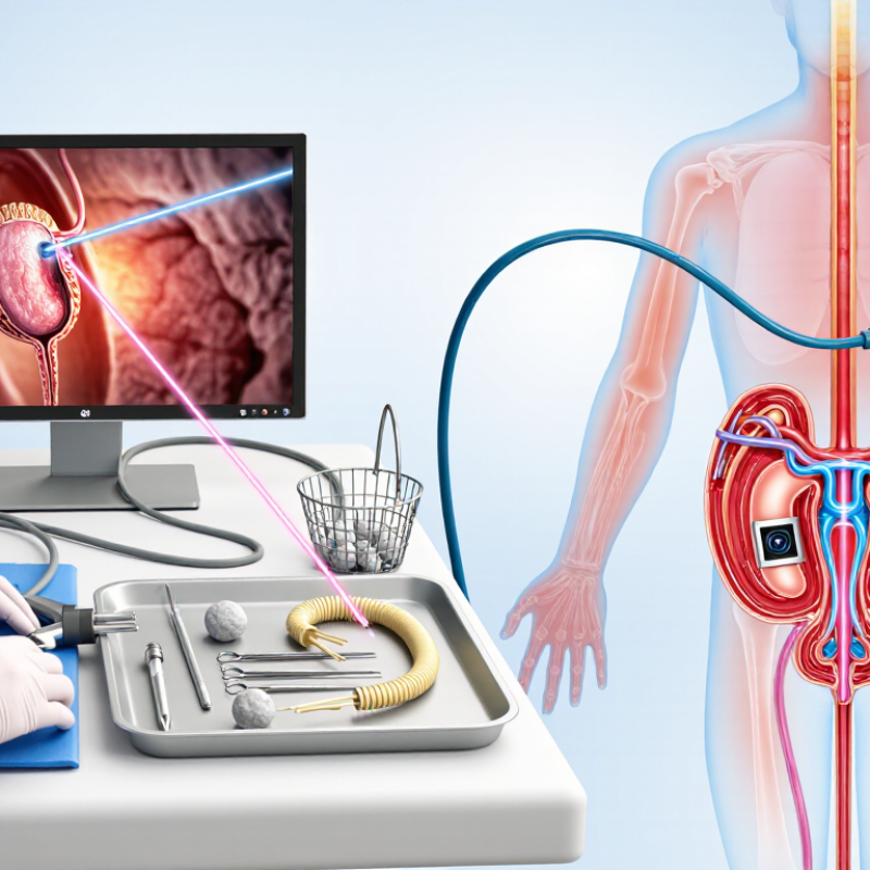
A ureteroscope is a thin instrument used to examine and treat the urinary tract. It passes through the urethra and bladder, then reaches the ureter. A small camera shows the stone on a monitor. The doctor may remove it with a basket or break it with laser energy. Flexible scopes can also reach stones inside the kidney.
Ureteroscopy is mainly used for ureteral and kidney stones that cause pain, blockage, bleeding, or repeated infections. It can also help investigate unusual narrowing or tissue changes. The National Institute of Diabetes and Digestive and Kidney Diseases reports that kidney stones affect about 11% of American men and 6% of women during their lifetimes. The European Association of Urology notes that treatment success depends on stone size, location, composition, and patient anatomy. Stone-free results are not guaranteed after one procedure.
The word “scope” sounds gentle. It is still surgery. Most patients receive anesthesia, and some need a temporary ureteral stent afterward. Possible effects include burning urination, blood in the urine, infection, or discomfort from the stent. Recovery varies more than many websites suggest.
Tips: Ask whether your stone needs removal or can pass naturally. Confirm the stone’s size and location. Discuss anesthesia, stent duration, infection precautions, and follow-up imaging. Bring questions; memory becomes unreliable under stress. Each case needs individualized advice from a qualified urologist.
A ureteroscope is a thin, lighted instrument for viewing the ureter and kidney. It can reveal stones, narrowed areas, bleeding, or unusual tissue.
The scope passes through the urethra and bladder into the ureter. Flexible models bend through natural curves using control wires and a hand lever.
A miniature camera sends close images to a monitor. Light fibers illuminate the area. Saline may wash away blood or particles.
Yes. A clinician may break a larger stone into smaller pieces using energy through the scope. A small basket can remove fragments.
Yes, when medically necessary. A thin tool can collect a small sample for laboratory testing.
Anesthesia is commonly used. You lie on your back while the clinician guides the scope gently. No external incision is usually needed.
No. A stent may support urine drainage while swelling settles. The care team should explain removal plans before discharge.
Mild burning, urinary urgency, or pink-tinged urine may occur. These effects can feel surprisingly noticeable. Recovery varies between patients.
Promptly report fever, worsening flank pain, heavy bleeding, or difficulty urinating. Small symptoms can matter. Follow-up confirms healing and normal urine flow.
A ureterscope is a slim, specialized instrument used to examine and treat the urinary tract, especially the ureters and kidneys. It may be flexible or rigid and includes a small viewing system, a light source, and channels that allow doctors to insert tiny instruments. These features help medical professionals identify stones, blockages, narrowing, bleeding, or other abnormalities without requiring a large incision.
During ureteroscopy, the instrument is carefully guided through the natural urinary passage into the bladder, ureter, or kidney. Doctors can inspect the area, remove or break up stones, collect tissue samples, and sometimes place a temporary stent to support urine flow. The procedure is commonly performed with anesthesia and is followed by monitoring and recovery instructions. Benefits may include accurate diagnosis, minimally invasive treatment, and a relatively short recovery period. Temporary discomfort, mild bleeding, urinary urgency, infection, or ureteral injury may occur, so patients should follow medical advice and report severe or persistent symptoms.